

The Real Cause of Heart Attacks: Understanding Atherosclerosis & Arterial Health
Cardiovascular disease kills more people every year than any other cause of death on earth. In the United States, someone dies of a heart attack or stroke every thirty-four seconds. Globally, cardiovascular disease accounts for approximately eighteen million deaths annually — roughly one in three of all deaths that occur on this planet.
At the centre of almost all of it is a process called atherosclerosis — the slow, decades-long accumulation of plaque inside artery walls that silently narrows blood vessels, hardens them, and eventually ruptures to cause the sudden, catastrophic events we call heart attacks and strokes.
And yet the public understanding of this process is almost entirely wrong.
Get health insights in your inbox
Weekly evidence-based tips on diabetes reversal and metabolic health. Unsubscribe anytime.
Most people have been taught that arterial plaque is what happens when you eat too much fat and cholesterol — that saturated fat raises LDL cholesterol, LDL deposits on artery walls like rust in a pipe, and eventually enough of it accumulates to block blood flow. The solution, in this picture, is to lower cholesterol — through diet, through statins, through reducing dietary fat — and the arteries will be protected.
This picture is not just incomplete. In its most important details, it is wrong in ways that have significant consequences for prevention and treatment.
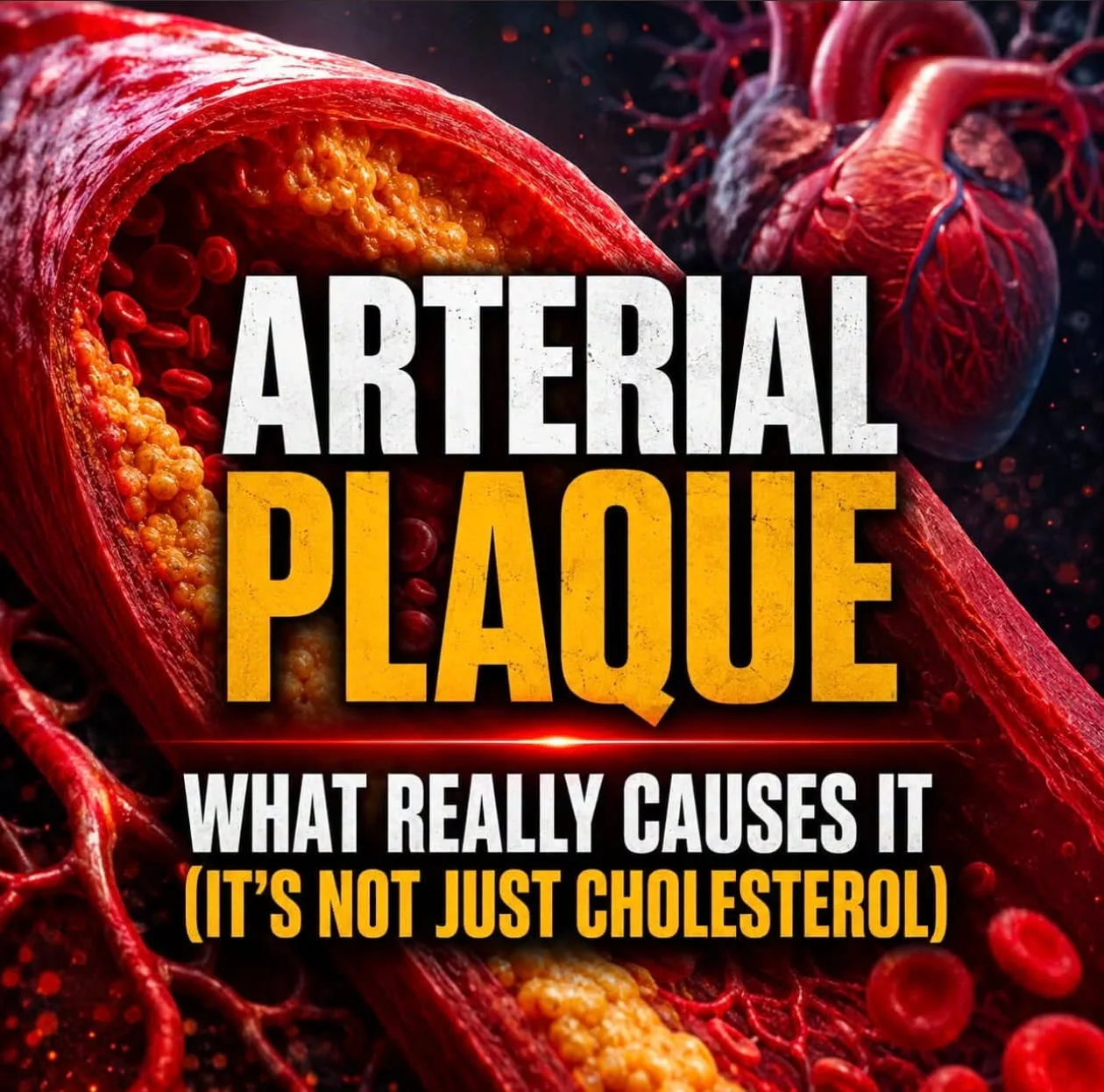
Arterial plaque is not a passive deposit of cholesterol on artery walls the way limescale builds in a pipe. It is an active, dynamic, inflammatory lesion — a wound response that begins not with cholesterol but with injury to the artery wall, that progresses not through cholesterol accumulation but through a chronic inflammatory cascade, that is driven by oxidised and modified lipoproteins rather than by cholesterol itself, and whose rupture — the event that actually causes heart attacks — is determined not by how large the plaque is but by how inflamed and unstable it is.
Cholesterol is involved. But it is involved in the way that white blood cells are involved in an infected wound — as a participant in a process whose underlying driver is something else entirely.
Understanding that underlying driver — and the complete network of dietary, lifestyle, metabolic, hormonal, and environmental factors that promote or prevent it — is what this guide is about.
🔬 𝗪𝗛𝗔𝗧 𝗔𝗥𝗧𝗘𝗥𝗜𝗔𝗟 𝗣𝗟𝗔𝗤𝗨𝗘 𝗔𝗖𝗧𝗨𝗔𝗟𝗟𝗬 𝗜𝗦
To understand plaque, you first need to understand what a healthy artery looks like — because plaque is not something that forms on the surface of arteries. It forms inside the artery wall itself.
𝗧𝗵𝗲 𝗮𝗿𝘁𝗲𝗿𝘆 𝘄𝗮𝗹𝗹
→ A healthy artery has three distinct layers. The innermost layer — the one that blood flows directly against — is called the endothelium. It is a single cell thick. One cell. This gossamer-thin lining is one of the most metabolically active tissues in the body: it regulates blood pressure through nitric oxide production, controls whether cells and molecules can pass into and out of the artery wall, prevents blood clotting under normal conditions, and acts as a selective barrier between the bloodstream and the underlying arterial tissue.
→ Beneath the endothelium is the middle layer — a thick band of smooth muscle cells embedded in a matrix of collagen and elastin. This layer gives arteries their mechanical strength and their ability to expand and contract with each heartbeat.
→ The outer layer provides structural support and houses the tiny blood vessels (vasa vasorum) that supply oxygen and nutrients to the artery wall itself.
→ Plaque does not build on the surface of arteries. It builds within the space between the endothelium and the smooth muscle layer — a space called the intima. The artery wall itself is where atherosclerosis lives.
𝗧𝗵𝗲 𝗮𝘁𝗵𝗲𝗿𝗼𝘀𝗰𝗹𝗲𝗿𝗼𝘁𝗶𝗰 𝗽𝗿𝗼𝗰𝗲𝘀𝘀 — 𝘀𝘁𝗲𝗽 𝗯𝘆 𝘀𝘁𝗲𝗽
𝗦𝘁𝗲𝗽 𝟭 — 𝗘𝗻𝗱𝗼𝘁𝗵𝗲𝗹𝗶𝗮𝗹 𝗶𝗻𝗷𝘂𝗿𝘆
→ Everything begins with damage to the endothelium. Something injures or irritates that single-cell lining — and the injured endothelium begins to malfunction. Instead of repelling immune cells and preventing inflammation, it starts producing adhesion molecules — molecular "velcro" that attracts white blood cells to the artery wall. Instead of producing nitric oxide that keeps the vessel relaxed and healthy, it produces inflammatory signals. Instead of acting as a selective barrier, it becomes leaky, allowing LDL particles to pass into the intima underneath.
→ What causes this endothelial injury? This is the crucial question — because it is the real beginning of the atherosclerotic process. The answer includes: oxidative stress from smoking, pollution, processed food chemicals, and excessive free radical production; chronic inflammation from any source — gut dysbiosis, metabolic syndrome, chronic infections, autoimmune conditions, psychological stress; mechanical stress from high blood pressure, which subjects the endothelium to abnormal physical forces with every heartbeat; high blood sugar and the glycation products it generates (AGEs — advanced glycation end-products) which directly damage endothelial cells; homocysteine excess, which is directly toxic to the endothelium; and excess insulin from insulin resistance, which promotes inflammation and oxidative stress throughout the arterial tree.
𝗦𝘁𝗲𝗽 𝟮 — 𝗟𝗗𝗟 𝗲𝗻𝘁𝗿𝘆 𝗮𝗻𝗱 𝗼𝘅𝗶𝗱𝗮𝘁𝗶𝗼𝗻
→ Once the endothelium is dysfunctional and leaky, LDL particles begin to enter the intima. But here is the critical point that the simple "cholesterol clogs pipes" story misses: LDL particles in the intima are not inherently dangerous. What makes them dangerous is what happens next — they get oxidised.
→ The intima, particularly in the context of inflammation and oxidative stress, is a chemically hostile environment. LDL particles that enter it are attacked by reactive oxygen species and modified — their cholesterol and fat content becomes oxidised, fundamentally changing their chemical character. Oxidised LDL (oxLDL) is not the same molecule as native LDL. It is inflammatory. It is toxic to endothelial cells. And critically, it is not recognised by the normal LDL receptor — it cannot be cleared through the usual cholesterol recycling pathway.
→ This is why the total LDL cholesterol number on a standard blood test tells an incomplete story. What matters is not simply how much LDL is present, but how much of it is being oxidised — and that depends on the oxidative stress environment of the artery wall, which depends on diet, lifestyle, metabolic health, and inflammatory burden.
𝗦𝘁𝗲𝗽 𝟯 — 𝗧𝗵𝗲 𝗶𝗺𝗺𝘂𝗻𝗲 𝗿𝗲𝘀𝗽𝗼𝗻𝘀𝗲 — 𝗳𝗼𝗮𝗺 𝗰𝗲𝗹𝗹𝘀 𝗮𝗻𝗱 𝘁𝗵𝗲 𝗳𝗮𝘁𝘁𝘆 𝘀𝘁𝗿𝗲𝗮𝗸
→ The injured endothelium releases chemical distress signals — MCP-1 (monocyte chemoattractant protein-1) and other chemokines — that recruit white blood cells called monocytes from the bloodstream into the intima. Once inside, monocytes differentiate into macrophages — the immune system's large clean-up cells, designed to engulf cellular debris and pathogens.
→ The macrophages encounter the oxidised LDL in the intima and begin to engulf it — their job is to clean up this dangerous modified material. But oxLDL is engulfed through a different receptor pathway (scavenger receptors rather than normal LDL receptors) — and this pathway has no feedback braking mechanism. Normal LDL uptake is regulated: when a cell has enough cholesterol, it downregulates its LDL receptors. Scavenger receptor uptake of oxLDL is unregulated — the macrophages keep engulfing oxLDL until they are so engorged with cholesterol and oxidised lipid that they transform into what pathologists call foam cells — bloated, lipid-filled cells that look foamy under the microscope.
→ These foam cells are the earliest visible lesion of atherosclerosis — the fatty streak. You can find fatty streaks in the aortas of children as young as ten years old in Western populations. The process begins this early, this silently.
𝗦𝘁𝗲𝗽 𝟰 — 𝗧𝗵𝗲 𝗶𝗻𝗳𝗹𝗮𝗺𝗺𝗮𝘁𝗼𝗿𝘆 𝗲𝘀𝗰𝗮𝗹𝗮𝘁𝗶𝗼𝗻
→ Foam cells do not just sit quietly. They release inflammatory cytokines — TNF-alpha, IL-1beta, IL-6 — that amplify the inflammatory response, recruit more immune cells, and cause smooth muscle cells from the middle layer of the artery to migrate into the intima and begin proliferating. These migrated smooth muscle cells produce collagen — beginning to build the fibrous cap that will eventually cover the growing plaque.
→ Some foam cells die in the intima, releasing their lipid contents into the extracellular space — forming what pathologists call the necrotic core: a pool of dead cells, oxidised lipid, cholesterol crystals, and cellular debris at the centre of the growing plaque.
→ More macrophages are recruited. More oxLDL is engulfed. More foam cells form and die. The necrotic core expands. T lymphocytes (another immune cell type) arrive and amplify the inflammatory signalling. The plaque grows — not like rust accumulating in a pipe, but like a chronic wound that the body keeps trying to heal but cannot quite resolve because the underlying drivers — the oxidative stress, the endothelial dysfunction, the metabolic dysfunction — are never removed.
𝗦𝘁𝗲𝗽 𝟱 — 𝗣𝗹𝗮𝗾𝘂𝗲 𝗺𝗮𝘁𝘂𝗿𝗮𝘁𝗶𝗼𝗻 𝗮𝗻𝗱 𝘁𝗵𝗲 𝗳𝗶𝗯𝗿𝗼𝘂𝘀 𝗰𝗮𝗽
→ As the plaque grows, smooth muscle cells continue producing collagen, forming a fibrous cap over the inflammatory core — essentially a scar tissue lid over the dangerous necrotic contents. A thick, well-organised fibrous cap is actually protective — it contains the dangerous material and prevents it from contacting the bloodstream.
→ Stable plaque has a thick fibrous cap and a relatively small necrotic core. It may narrow the artery significantly but it is less likely to rupture acutely. Stable plaque can cause symptoms (angina, claudication) from reduced blood flow, and is addressable with arterial bypass or stenting when flow limitation is severe.
→ Vulnerable plaque — the genuinely dangerous lesion — has a thin, weakened fibrous cap, a large necrotic core, and active inflammation at its shoulders (the edges where the plaque meets normal artery wall). Vulnerable plaque may cause little or no blood flow limitation — it may not even show up as significant narrowing on a coronary angiogram. But it is primed to rupture.
𝗦𝘁𝗲𝗽 𝟲 — 𝗣𝗹𝗮𝗾𝘂𝗲 𝗿𝘂𝗽𝘁𝘂𝗿𝗲 — 𝘄𝗵𝗮𝘁 𝗮𝗰𝘁𝘂𝗮𝗹𝗹𝘆 𝗰𝗮𝘂𝘀𝗲𝘀 𝗵𝗲𝗮𝗿𝘁 𝗮𝘁𝘁𝗮𝗰𝗸𝘀
→ When a vulnerable plaque ruptures — when that thin fibrous cap tears — the necrotic core material is suddenly exposed to the flowing bloodstream. The body interprets this as a catastrophic wound. The clotting cascade activates instantly and massively. A blood clot (thrombus) forms on the ruptured plaque surface within seconds to minutes.
→ If that clot is large enough to completely obstruct the coronary artery, the heart muscle downstream receives no oxygen — this is a myocardial infarction. If the clot travels upstream to the brain, it is a stroke. If the clot partially obstructs the artery, it produces the chest pain syndrome called unstable angina.
→ This is the critical clinical insight: most fatal heart attacks do not occur in arteries that were severely narrowed. Post-mortem studies and angiographic studies consistently show that most heart attack-causing plaques were causing less than 50% arterial narrowing before they ruptured — they were not the plaques that looked dangerous on imaging. The danger was not in their size but in their instability — in the activity of the inflammatory process inside them.
→ Plaque stability — not plaque size — is the most important determinant of heart attack risk. And plaque stability is determined by the balance between inflammation (which thins the fibrous cap through matrix metalloproteinase activity) and anti-inflammatory, plaque-stabilising processes (which thicken the cap and reduce necrotic core activity).
This is why treating atherosclerosis purely as a cholesterol management problem — reducing LDL and calling it done — addresses only one part of the picture. It does not address endothelial function, oxidative stress, inflammation, plaque stability, or any of the upstream metabolic drivers that determine whether plaque forms and whether it ruptures.
🩸 𝗖𝗛𝗢𝗟𝗘𝗦𝗧𝗘𝗥𝗢𝗟 — 𝗧𝗛𝗘 𝗛𝗢𝗡𝗘𝗦𝗧 𝗣𝗜𝗖𝗧𝗨𝗥𝗘
Cholesterol is genuinely important to the atherosclerotic process. But the public understanding of it — framed entirely as "high cholesterol causes heart disease, lower it with diet and drugs" — misses most of what the science actually shows.
→ Cholesterol is not a toxin. It is an essential molecule — a structural component of every cell membrane in the body; the precursor for all steroid hormones (cortisol, oestrogen, testosterone, progesterone, DHEA); the precursor for vitamin D; the precursor for bile acids; and a major component of myelin, the insulating sheath around nerve fibres. The brain is approximately 25% cholesterol by dry weight. Your body produces roughly 80% of your cholesterol endogenously regardless of dietary intake — because it is too important to leave entirely to diet.
→ LDL is not inherently bad. LDL (low-density lipoprotein) is a transport vehicle — it carries cholesterol and fat-soluble nutrients from the liver to every cell in the body that needs them. Every cell uses LDL cholesterol for membrane maintenance, hormone production, and vitamin D synthesis. Without LDL, cells cannot function.
→ What matters is not simply how much LDL is present but the characteristics of the LDL particles. Small, dense LDL particles are far more atherogenic than large, buoyant LDL particles — small dense particles penetrate the endothelium more easily, are more susceptible to oxidation, and are cleared more slowly. A person with a high total LDL number but predominantly large buoyant particles may have lower actual cardiovascular risk than someone with a lower LDL number but predominantly small dense particles. The standard total LDL cholesterol test does not tell you which type predominates.
→ Oxidised LDL is the actual culprit. As described above, it is not native LDL but oxidised LDL that is engulfed by macrophages, forms foam cells, and drives the atherosclerotic cascade. The key risk factor is not LDL level per se but the oxidative environment in which LDL exists — which is determined by antioxidant status, metabolic health, dietary quality, and inflammatory burden.
→ HDL does far more than people realise. HDL (high-density lipoprotein) is the cholesterol transport vehicle that picks up excess cholesterol from peripheral tissues — including from early atherosclerotic lesions — and returns it to the liver for reprocessing or excretion. This reverse cholesterol transport is one of the most important protective mechanisms against plaque progression. But HDL also has direct anti-inflammatory effects on the endothelium, anti-oxidant properties, and direct efflux-promoting activity on foam cells. Low HDL is a stronger independent predictor of cardiovascular events than high LDL in many population studies.
→ Triglycerides matter enormously and are underemphasised. High blood triglycerides — driven primarily by refined carbohydrates and sugar rather than dietary fat — promote the formation of small dense LDL, reduce HDL, and contribute directly to endothelial inflammation. The triglyceride:HDL ratio is one of the most powerful and most underutilised markers of cardiovascular risk available on a standard lipid panel. A triglyceride:HDL ratio above 2 (in mg/dL units) or above 0.9 (in mmol/L units) is a strong indicator of insulin resistance and small dense LDL predominance — far more predictive of cardiovascular risk in many populations than LDL alone.
→ Lipoprotein(a) — the risk factor medicine mostly ignores. Lp(a) is a specific type of LDL particle with an additional protein called apolipoprotein(a) attached. It is far more atherogenic than regular LDL — it promotes both atherosclerosis and thrombosis (blood clotting). Levels are almost entirely genetically determined and are largely unaffected by diet and lifestyle. Approximately 20% of the population has elevated Lp(a). It is not measured on a standard lipid panel. And it is one of the most important independent cardiovascular risk factors identified — people with elevated Lp(a) can have normal or even low standard LDL while carrying significantly elevated cardiovascular risk. Lp(a) testing should be routine and is not.
→ ApoB is more informative than LDL-C. ApoB (apolipoprotein) - is a protein found on every atherogenic lipoprotein particle — LDL, VLDL, IDL, and Lp(a). Each particle carries exactly one ApoB molecule. ApoB directly measures the total number of atherogenic particles — which is what actually matters, not the total amount of cholesterol they carry. Two people can have the same LDL cholesterol number but one has it carried in fewer, larger particles (lower ApoB, lower risk) and the other in more, smaller particles (higher ApoB, higher risk). ApoB is the more accurate atherosclerotic risk marker.
🔥 𝗪𝗛𝗔𝗧 𝗔𝗖𝗧𝗨𝗔𝗟𝗟𝗬 𝗗𝗥𝗜𝗩𝗘𝗦 𝗣𝗟𝗔𝗤𝗨𝗘 — 𝗧𝗛𝗘 𝗖𝗢𝗠𝗣𝗟𝗘𝗧𝗘 𝗣𝗜𝗖𝗧𝗨𝗥𝗘
𝗜𝗻𝘀𝘂𝗹𝗶𝗻 𝗿𝗲𝘀𝗶𝘀𝘁𝗮𝗻𝗰𝗲 — 𝘁𝗵𝗲 𝗺𝗼𝘀𝘁 𝗶𝗺𝗽𝗼𝗿𝘁𝗮𝗻𝘁 𝗺𝗼𝗱𝗶𝗳𝗶𝗮𝗯𝗹𝗲 𝗿𝗶𝘀𝗸 𝗳𝗮𝗰𝘁𝗼𝗿
→ Insulin resistance — the state in which cells stop responding normally to insulin, requiring the pancreas to produce ever-higher insulin levels to maintain blood sugar — is now understood to be the central metabolic driver of atherosclerosis in most people in the modern world.
→ High circulating insulin directly promotes atherosclerosis through multiple pathways: it stimulates smooth muscle cell proliferation in the artery wall; it promotes endothelial inflammation; it increases VLDL and triglyceride production; it shifts LDL toward the small dense atherogenic pattern; it raises blood pressure; and it promotes a prothrombotic state — more likely to form dangerous blood clots on ruptured plaques.
→ People with metabolic syndrome — the cluster of insulin resistance, central obesity, elevated triglycerides, low HDL, elevated blood pressure, and elevated fasting glucose — have three to five times the cardiovascular event risk of metabolically healthy people. This cluster is present in approximately one third of adults in most Western countries.
→ Critically: people with insulin resistance and metabolic syndrome may have completely normal or even low LDL cholesterol while having severely atherogenic lipid profiles — high triglycerides, low HDL, and small dense LDL. They may be told their cholesterol is fine while the actual metabolic drivers of their plaque are completely unaddressed.
𝗛𝘆𝗽𝗲𝗿𝘁𝗲𝗻𝘀𝗶𝗼𝗻
→ High blood pressure exerts physical shear stress on the endothelium with every heartbeat. The turbulent blood flow at arterial branch points — where blood diverts and changes direction — creates particularly intense mechanical stress on the endothelial lining. This is precisely why atherosclerotic plaques preferentially form at arterial branch points: the coronary artery branches, the carotid bifurcation, the aortic bifurcation, the iliac bifurcation. High blood pressure intensifies this mechanical endothelial injury at every vulnerable point in the arterial tree with every pulse.
→ Every 20 mmHg increase in systolic blood pressure approximately doubles the risk of cardiovascular death. Optimal blood pressure for arterial health is below 120/80 mmHg — not the 140/90 that is conventionally defined as the treatment threshold.
𝗖𝗵𝗿𝗼𝗻𝗶𝗰 𝗶𝗻𝗳𝗹𝗮𝗺𝗺𝗮𝘁𝗶𝗼𝗻
→ Systemic inflammation amplifies every step of the atherosclerotic cascade — it promotes endothelial dysfunction, increases LDL oxidation, promotes foam cell formation, thins the fibrous cap through matrix metalloproteinase activation, and creates the pro-thrombotic environment that determines whether plaque rupture leads to full arterial occlusion.
→ hs-CRP (high-sensitivity C-reactive protein) — the standard clinical inflammatory marker — is an independent predictor of cardiovascular events even in people with normal or low LDL. The JUPITER trial demonstrated that people with elevated hs-CRP but normal LDL levels benefited from statin therapy through its anti-inflammatory effects — not (or not only) through its LDL-lowering effects.
→ Sources of chronic low-grade inflammation relevant to cardiovascular risk include: gut dysbiosis and intestinal permeability ("leaky gut"); periodontal (gum) disease — one of the most underappreciated cardiovascular risk factors with some of the strongest epidemiological evidence; chronic psychological stress and depression; sleep deprivation; visceral adiposity (fat around the abdominal organs is a major source of inflammatory cytokines); chronic infections (particularly Chlamydophila pneumoniae and Helicobacter pylori have been associated with plaque progression); air pollution; and the chronic low-grade endotoxaemia from gut bacterial products entering the bloodstream through a compromised intestinal barrier.
𝗢𝘅𝗶𝗱𝗮𝘁𝗶𝘃𝗲 𝘀𝘁𝗿𝗲𝘀𝘀
→ Oxidative stress — the imbalance between free radical production and antioxidant defences — is the immediate chemical mechanism driving LDL oxidation in the artery wall. Without LDL oxidation, LDL particles in the intima are not atherogenic. With high oxidative stress, even modest LDL concentrations can be extensively oxidised and highly atherogenic.
→ Sources of vascular oxidative stress: smoking (the single most powerful external oxidative stressor on the vasculature); processed seed oils high in omega-6 linoleic acid, which are highly susceptible to peroxidation; dietary advanced glycation end-products (AGEs) from high-heat processed foods; high blood sugar (glucose directly generates reactive oxygen species); excess iron (iron catalyses free radical reactions); pollution exposure; heavy metal accumulation (lead, cadmium, arsenic); and inadequate dietary antioxidants.
𝗛𝗼𝗺𝗼𝗰𝘆𝘀𝘁𝗲𝗶𝗻𝗲
→ Homocysteine is an amino acid produced as a byproduct of methionine metabolism. At elevated levels, it is directly toxic to endothelial cells — it increases oxidative stress in the artery wall, impairs nitric oxide production, promotes smooth muscle cell proliferation, and creates a pro-thrombotic arterial environment.
→ Elevated homocysteine is a significant independent cardiovascular risk factor. And it is almost entirely caused by nutritional deficiencies — specifically inadequate folate, vitamin B6, and vitamin B12, which are the cofactors required for the enzymes that clear homocysteine from the body. The MTHFR gene variant also impairs homocysteine clearance and is present in a significant proportion of the population.
→ Homocysteine is not measured on a standard cardiovascular risk panel. It should be.
𝗛𝗶𝗴𝗵 𝗯𝗹𝗼𝗼𝗱 𝘀𝘂𝗴𝗮𝗿 𝗮𝗻𝗱 𝗔𝗚𝗘𝘀
→ Chronically elevated blood glucose directly damages endothelial cells through multiple mechanisms — generating reactive oxygen species, activating inflammatory pathways (NF-kB), and producing advanced glycation end-products (AGEs).
→ AGEs are compounds formed when sugar molecules bond chemically to proteins or fats — "caramelising" them and impairing their function. In the artery wall, AGEs cross-link collagen fibres, making arteries stiffer. They directly activate inflammatory receptors (RAGE — receptor for advanced glycation end-products) on endothelial cells and macrophages — amplifying the inflammatory cascade inside the plaque. People with diabetes and prediabetes have dramatically accelerated plaque progression for exactly these reasons.
→ Dietary AGEs — formed when food is cooked at high temperatures through processes like frying, grilling, and browning — add to the total AGE load independently of blood sugar levels. The AGE content of the modern diet has increased dramatically as processed and ultra-processed food consumption has risen.
𝗦𝗺𝗼𝗸𝗶𝗻𝗴
→ Smoking is the most powerful single modifiable cardiovascular risk factor. It damages the endothelium through direct chemical toxicity and oxidative stress; dramatically increases LDL oxidation; reduces HDL; increases fibrinogen (a clotting protein — promoting the thrombosis that makes plaque rupture lethal); promotes inflammation; and increases blood pressure. Smokers have two to four times the heart attack risk of non-smokers, and the risk is dose-dependent. Within one year of stopping, cardiovascular risk begins to fall significantly.
𝗦𝗹𝗲𝗲𝗽 𝗱𝗲𝗽𝗿𝗶𝘃𝗮𝘁𝗶𝗼𝗻
→ Consistently sleeping less than six hours per night is associated with a two- to three-fold increase in cardiovascular events in prospective studies. The mechanisms are multiple: sleep deprivation raises cortisol (promoting endothelial inflammation and insulin resistance); raises blood pressure; reduces HRV (heart rate variability — a marker of autonomic nervous system health); increases inflammatory markers; impairs glucose tolerance; and reduces the overnight endothelial repair processes that maintain arterial wall integrity. Sleep is not optional for arterial health.
𝗖𝗵𝗿𝗼𝗻𝗶𝗰 𝘀𝘁𝗿𝗲𝘀𝘀
→ Chronic psychological stress activates the hypothalamic-pituitary-adrenal axis, producing sustained cortisol elevation, and the sympathetic nervous system, producing sustained adrenaline and noradrenaline elevation. These stress hormones raise blood pressure, promote endothelial inflammation, shift lipid profiles toward the atherogenic pattern, promote platelet aggregation (increasing clotting tendency), impair sleep, and promote visceral fat accumulation — through every one of these mechanisms, chronic stress accelerates atherosclerotic progression.
→ Depression independently doubles cardiovascular event risk — through inflammation, reduced HRV, reduced physical activity, poorer dietary choices, and direct neurobiological effects on platelet function. The cardiovascular consequences of mental health cannot be separated from the physical ones.
𝗢𝗿𝗮𝗹 𝗵𝗲𝗮𝗹𝘁𝗵
→ Periodontal disease — chronic bacterial infection of the gums — is one of the most striking and most underappreciated cardiovascular risk factors. The same bacteria responsible for gum disease (Porphyromonas gingivalis, Streptococcus sanguis) have been found inside coronary artery plaques. Chronic gum infection generates persistent systemic inflammation, elevates fibrinogen and hs-CRP, and introduces pro-inflammatory bacterial products into the bloodstream with every chewing episode.
→ Multiple large prospective studies show that people with periodontal disease have significantly higher rates of heart attack and stroke, independent of traditional risk factors. Treating periodontal disease reduces systemic inflammatory markers. Flossing, gum health maintenance, and regular dental care are cardiovascular interventions — not just cosmetic ones.
🌿 𝗧𝗛𝗘 𝗖𝗢𝗠𝗣𝗟𝗘𝗧𝗘 𝗜𝗡𝗧𝗘𝗥𝗩𝗘𝗡𝗧𝗜𝗢𝗡 𝗣𝗥𝗢𝗧𝗢𝗖𝗢𝗟
𝗗𝗶𝗲𝘁 — 𝘁𝗵𝗲 𝗳𝗼𝘂𝗻𝗱𝗮𝘁𝗶𝗼𝗻
→ The Mediterranean dietary pattern has the strongest evidence base of any dietary pattern for cardiovascular event reduction — not just risk factor modification, but actual heart attacks and strokes prevented. The landmark PREDIMED trial (over 7,000 high-risk adults followed for five years) found the Mediterranean diet supplemented with extra-virgin olive oil or nuts reduced major cardiovascular events by approximately 30% compared to a low-fat control diet. No medication trial for primary cardiovascular prevention has produced comparable results. The core of this diet: abundant olive oil, vegetables, legumes, whole grains, fish, nuts, and moderate whole fruit — with red meat, processed food, sugar-sweetened beverages, and refined grains minimised.
→ Eliminate refined carbohydrates and added sugars — not primarily because of their caloric content but because of their metabolic effects. Refined carbohydrates drive the hyperinsulinaemia that promotes small dense LDL, raises triglycerides, lowers HDL, increases visceral adiposity, promotes endothelial inflammation, and drives the entire insulin resistance-atherosclerosis cascade. Lowering dietary glycaemic load is one of the most powerful lipid-improving, inflammation-reducing, and insulin-sensitising interventions available.
→ Extra-virgin olive oil is the single most cardiovascular-protective dietary fat. Its monounsaturated oleic acid is highly resistant to oxidation — reducing the lipid peroxidation products that damage endothelial cells and oxidise LDL. Its polyphenols (particularly oleocanthal and oleuropein) have direct anti-inflammatory effects — oleocanthal has been shown to inhibit the same enzyme (COX) as ibuprofen, through a different binding mechanism. EVOO specifically has been shown to improve endothelial function, reduce LDL oxidation, improve HDL functionality, and reduce platelet aggregation. The target: 3–4 tablespoons of high-quality EVOO daily.
→ Eliminate processed seed oils — refined corn, soybean, sunflower, safflower, cottonseed, and canola oils. These oils are extremely high in omega-6 linoleic acid, which is highly susceptible to peroxidation at cooking temperatures. Oxidised linoleic acid metabolites (OXLAMs) are directly pro-inflammatory, directly toxic to endothelial cells, and may drive the oxidation of LDL particles in the artery wall. The omega-6 to omega-3 ratio in the Western diet has shifted from an ancestral approximately 4:1 to a modern approximately 20:1 — a shift that fundamentally alters the inflammatory baseline of the vascular system.
→ Increase omega-3 fatty acids. EPA and DHA from oily fish and marine sources reduce triglycerides, reduce platelet aggregation, reduce inflammation (EPA is the precursor to anti-inflammatory eicosanoids), improve endothelial function, stabilise the electrical activity of the heart (reducing arrhythmia risk), and — at higher doses — may directly reduce cardiovascular events. Multiple large trials including REDUCE-IT and STRENGTH have examined high-dose omega-3 supplementation with mixed but overall supportive results. The food-based recommendation: oily fish (sardines, mackerel, wild salmon, herring, anchovies) at least three times weekly. Supplement dose for cardiovascular support: 2–4g EPA+DHA daily.
→ Increase dietary nitrates from leafy greens and beetroot. Dietary nitrates are converted in the body to nitric oxide — the molecule the endothelium produces to maintain arterial relaxation, prevent platelet adhesion, and protect against endothelial dysfunction. Dark leafy greens (arugula/rocket has the highest nitrate content, followed by spinach, kale, and Swiss chard) and beetroot are the most concentrated dietary nitrate sources. Consuming them consistently is one of the most evidence-supported dietary strategies for lowering blood pressure and improving endothelial function.
→ Berries — particularly blueberries, strawberries, and pomegranate — contain anthocyanins and other polyphenols that have direct endothelial-protective effects. The Nurses' Health Study found that women consuming three or more servings of blueberries and strawberries weekly had a 34% lower heart attack risk than those consuming less — a finding that has been supported by multiple mechanistic studies showing improved endothelial function, reduced oxLDL, and reduced inflammatory markers from regular berry consumption.
→ Fibre — particularly soluble fibre from oats, legumes, flaxseed, and vegetables — binds bile acids in the gut, stimulating the liver to produce more bile acids from LDL cholesterol, effectively reducing circulating LDL. The beta-glucan in oats has the most consistent evidence for LDL reduction — approximately 5–10% reduction with 3g beta-glucan daily. Fibre also feeds the gut microbiome that produces short-chain fatty acids protective against arterial inflammation, and reduces post-meal blood sugar spikes that drive glycation and oxidative stress.
→ Garlic has direct anti-atherosclerotic effects through multiple mechanisms — it reduces LDL oxidation, reduces platelet aggregation (its ajoene and thiosulfinates directly inhibit platelet activation), lowers blood pressure through nitric oxide-potentiating effects, mildly lowers LDL, and has anti-inflammatory activity. Aged garlic extract (AGE) has specific evidence for reducing coronary artery calcification progression in a randomised controlled trial. A 2016 study by Budoff and colleagues found that aged garlic extract reduced the progression of coronary artery calcium score significantly over one year compared to placebo. Dose: 600–1,200mg aged garlic extract daily, or 2–4 raw garlic cloves daily.
→ Dark chocolate and cocoa — high-flavanol cocoa has documented endothelial benefits: increasing nitric oxide production, reducing blood pressure, reducing LDL oxidation, and reducing platelet aggregation. The evidence specifically supports high-flavanol cocoa (not commercial low-flavanol chocolate), but high-quality dark chocolate (85%+ cocoa) retains meaningful flavanol content. The dose in the evidence: 20–40g dark chocolate or 200–400mg cocoa flavanols daily.
𝗞𝗲𝘆 𝗻𝘂𝘁𝗿𝗶𝗲𝗻𝘁𝘀 𝗮𝗻𝗱 𝘀𝘂𝗽𝗽𝗹𝗲𝗺𝗲𝗻𝘁𝘀
→ Vitamin K2 (MK-7) is one of the most important and most underappreciated vascular nutrients. As introduced in the bone health guide — Matrix Gla Protein (MGP) is the body's primary inhibitor of arterial calcification. It must be carboxylated (activated) by vitamin K2 to function. Without adequate K2, MGP sits inactive, and calcium accumulates in artery walls — producing arterial calcification, the stiffening of arteries that dramatically increases cardiovascular risk. Arterial calcification is measured by coronary artery calcium (CAC) scoring — currently the most powerful imaging predictor of cardiovascular events available. The Rotterdam Study found that the highest vitamin K2 intake was associated with a 57% reduction in coronary heart disease mortality and significantly less aortic calcification. Dose: MK-7 180mcg daily with a fat-containing meal.
→ Magnesium is required for over 300 enzymatic reactions in the body, including those that regulate blood pressure, maintain endothelial function, prevent arterial spasm, and regulate the electrical activity of the heart. Magnesium deficiency is associated with hypertension, endothelial dysfunction, elevated inflammatory markers, and increased risk of arrhythmia. Large epidemiological studies consistently show that higher dietary magnesium is associated with lower cardiovascular mortality. Magnesium also relaxes smooth muscle — contributing directly to blood pressure reduction. Dose: 300–400mg magnesium glycinate or malate daily.
→ Vitamin D deficiency is independently associated with cardiovascular disease in multiple large prospective studies. Vitamin D has direct effects on the vascular system: it reduces inflammatory cytokine production, promotes endothelial function, reduces smooth muscle cell proliferation in the artery wall, reduces parathyroid hormone (which promotes vascular calcification when elevated), and modulates the renin-angiotensin system that regulates blood pressure. While large RCTs of vitamin D supplementation have not consistently shown cardiovascular event reduction — likely because the trials enrolled people with already adequate levels or used insufficient doses — the mechanistic and epidemiological evidence supports maintaining optimal vitamin D status (75–125 nmol/L) as a component of cardiovascular health. Dose: 2,000–5,000 IU vitamin D3 daily.
→ CoQ10 (ubiquinol) is essential for mitochondrial energy production in heart muscle cells — the heart has the highest CoQ10 content of any organ because of its continuous energy demands. CoQ10 is also a fat-soluble antioxidant that specifically protects LDL from oxidation in the bloodstream. Statin medications deplete CoQ10 by inhibiting the same mevalonate pathway that produces both cholesterol and CoQ10 — everyone on statins should be supplementing CoQ10. Beyond statins: CoQ10 has clinical evidence for reducing blood pressure and improving endothelial function. Dose: 200–400mg ubiquinol (the reduced, more bioavailable form) daily.
→ Nattokinase is a fibrinolytic enzyme extracted from natto (fermented soybeans) — it breaks down fibrin, the primary structural protein of blood clots, through a mechanism similar to but distinct from pharmaceutical thrombolytics. Multiple clinical studies have shown nattokinase reduces blood clotting tendency, reduces fibrinogen levels, and in some studies modestly reduces LDL and blood pressure. For people concerned about thrombotic risk (the clot that forms on a ruptured plaque is what makes heart attacks lethal), nattokinase provides natural fibrinolytic support. Important: avoid in people already on anticoagulants without medical supervision; avoid before surgery. Dose: 2,000–4,000 FU (fibrinolytic units) daily, taken away from food.
→ Bergamot (from Bergamot orange extract) has emerged as one of the most evidence-supported botanical interventions for lipid management. Multiple clinical trials in Italy — where bergamot has been used as a cholesterol-lowering supplement for decades — have demonstrated significant reductions in LDL, triglycerides, and oxLDL alongside increases in HDL. The polyphenols in bergamot activate AMPK (the same metabolic master switch activated by exercise and metformin), inhibit HMG-CoA reductase (the same enzyme targeted by statins, but through a different mechanism), and specifically reduce oxLDL — addressing the oxidised LDL that is the actual atherogenic species. Dose: 500–1,000mg standardised bergamot extract daily.
→ Folate, B6, and B12 for homocysteine clearance. Elevated homocysteine is a direct endothelial toxin and an independent cardiovascular risk factor — and it is almost entirely nutritionally driven. Adequate folate (or methylfolate for those with MTHFR variants), vitamin B6, and vitamin B12 are the cofactors required for the enzymes that clear homocysteine. B12 is particularly important for the elderly and for anyone on metformin (which reduces B12 absorption). Testing homocysteine and addressing elevation with targeted B vitamin supplementation is one of the most straightforward and underutilised cardiovascular risk reductions available. Target homocysteine: below 10 µmol/L; optimal below 7 µmol/L. Dose: methylfolate 400–800mcg daily; B6 (P-5-P form) 25–50mg daily; B12 (methylcobalamin) 500–1,000mcg daily.
→ Berberine has extensive clinical evidence as a metabolic and cardiovascular agent — it activates AMPK, significantly reduces LDL (approximately 20–25% in multiple trials), reduces triglycerides, reduces blood sugar, reduces insulin resistance, and has direct anti-inflammatory and anti-atherosclerotic effects in animal models. For people with metabolic syndrome, elevated LDL, or insulin resistance seeking a non-statin lipid intervention, berberine is the most evidence-supported botanical option. Dose: 500mg three times daily with meals (berberine has a short half-life requiring multiple daily doses for sustained effect).
→ Resveratrol and pterostilbene — the polyphenols from red grapes and blueberries respectively — have direct evidence for improving endothelial function, reducing LDL oxidation, reducing inflammatory markers, improving insulin sensitivity, and activating SIRT1 (a longevity-associated gene that improves vascular function). Pterostilbene has superior bioavailability to resveratrol and is increasingly preferred. Dose: resveratrol 150–500mg daily; pterostilbene 50–250mg daily.
→ Vitamin C has specific endothelial-regenerating effects — it is a required cofactor for collagen synthesis (maintaining arterial wall structural integrity), a direct antioxidant that protects LDL from oxidation, and a nitric oxide-sparing agent that improves endothelial function. Multiple clinical studies show vitamin C supplementation improves endothelial function as measured by flow-mediated dilation. Dose: 500–1,000mg twice daily (food sources alongside supplementation: bell peppers, kiwi, citrus, broccoli, strawberries).
𝗘𝘅𝗲𝗿𝗰𝗶𝘀𝗲 — 𝘁𝗵𝗲 𝗺𝗼𝘀𝘁 𝗽𝗼𝘄𝗲𝗿𝗳𝘂𝗹 𝘀𝗶𝗻𝗴𝗹𝗲 𝗮𝗻𝘁𝗶-𝗮𝘁𝗵𝗲𝗿𝗼𝘀𝗰𝗹𝗲𝗿𝗼𝘁𝗶𝗰 𝗶𝗻𝘁𝗲𝗿𝘃𝗲𝗻𝘁𝗶𝗼𝗻
→ Regular aerobic exercise is the most comprehensively anti-atherosclerotic intervention available — it improves every major driver of plaque simultaneously. It increases HDL; reduces triglycerides; shifts LDL toward the larger, less atherogenic pattern; reduces blood pressure; improves insulin sensitivity; reduces visceral adiposity; reduces systemic inflammation (regular exercise chronically lowers hs-CRP); improves endothelial function through increased nitric oxide production in response to blood flow shear stress; and reduces sympathetic nervous system tone (lowering resting heart rate, reducing the mechanical stress on the endothelium with every heartbeat).
→ Zone 2 aerobic exercise — the intensity at which you can sustain a conversation but feel meaningfully challenged, approximately 60–70% of maximum heart rate — is the most important training zone for cardiovascular and metabolic health. At this intensity, mitochondrial biogenesis is maximally stimulated, fat oxidation is prioritised, and parasympathetic tone is supported. Target: 150–300 minutes of Zone 2 exercise weekly.
→ High-intensity interval training (HIIT) provides additional cardiovascular benefits beyond Zone 2 — particularly for VO2max (maximal oxygen uptake), which is one of the strongest predictors of cardiovascular mortality available. Even two to three short HIIT sessions weekly (4–6 rounds of high-intensity effort with recovery intervals) produces significant VO2max improvements. VO2max improvement is associated with reduced all-cause mortality independently of other risk factors.
→ Resistance training two to three times weekly adds independent cardiovascular benefit through insulin sensitisation, reduced visceral adiposity, improved glucose metabolism, and direct blood pressure lowering effects. The combination of aerobic and resistance training is superior to either alone.
→ Reducing sedentary time matters independently of exercise. Sitting for prolonged uninterrupted periods increases cardiovascular risk even in people who exercise regularly — the mechanisms include reduced lipoprotein lipase activity (impairing triglyceride clearance), reduced glucose uptake in resting muscles, and increased clotting tendency. Breaking prolonged sitting with brief movement every 30–60 minutes meaningfully reduces these effects.
𝗟𝗶𝗳𝗲𝘀𝘁𝘆𝗹𝗲 𝗳𝗮𝗰𝘁𝗼𝗿𝘀
→ Stop smoking — the single most impactful lifestyle cardiovascular intervention available. Within one year of stopping, excess heart attack risk falls by approximately 50%. Within fifteen years, cardiovascular risk approaches that of a non-smoker. No other intervention produces this magnitude of benefit in this timeframe.
→ Prioritise sleep — seven to nine hours of quality sleep per night. Sleep deprivation is not a minor contributor; it is a major, underappreciated cardiovascular risk factor. Establishing a consistent sleep schedule, reducing blue light exposure in the evening, keeping the bedroom cool and dark, and addressing sleep apnoea (which independently drives cardiovascular risk through nocturnal hypoxia and inflammatory activation) are all legitimate cardiovascular health interventions.
→ Manage stress — not as a lifestyle nicety but as a specific cardiovascular prescription. The parasympathetic practices discussed in the polyvagal theory and nervous system regulation guides — coherent breathing (particularly slow diaphragmatic breathing at 5–6 breaths per minute, which directly activates the baroreflex and restores heart rate variability), nature exposure, social connection, and meditation — produce measurable reductions in cortisol, blood pressure, inflammatory markers, and platelet activation. The Transcendental Meditation literature has particularly strong cardiovascular evidence, including a meta-analysis showing significant blood pressure reduction from regular meditation practice.
→ Maintain oral health — floss daily, brush twice daily, see a dentist regularly, and take periodontal disease seriously as the cardiovascular risk factor it is. The bacteria in diseased gums have been found in coronary plaques. The inflammatory burden of active gum disease is cardiovascularly measurable. Treating periodontal disease reduces systemic hs-CRP and fibrinogen.
→ Minimise toxin exposure — air pollution, cigarette smoke (including second-hand), and heavy metals (particularly lead and cadmium from smoking and certain foods) all directly promote endothelial damage and LDL oxidation. Regular sauna use has specific evidence for cardiovascular benefit — the Finnish sauna studies showed dramatically lower cardiovascular mortality in men who used a sauna four or more times weekly, through mechanisms including improved endothelial function, blood pressure reduction, improved autonomic nervous system balance, and direct heat-stress conditioning of the vasculature.
🔬 𝗪𝗛𝗔𝗧 𝗧𝗢 𝗠𝗘𝗔𝗦𝗨𝗥𝗘 — 𝗕𝗘𝗬𝗢𝗡𝗗 𝗧𝗛𝗘 𝗦𝗧𝗔𝗡𝗗𝗔𝗥𝗗 𝗖𝗛𝗢𝗟𝗘𝗦𝗧𝗘𝗥𝗢𝗟 𝗣𝗔𝗡𝗘𝗟
A standard lipid panel (total cholesterol, LDL, HDL, triglycerides) provides a partial and often misleading picture of cardiovascular risk. A more complete assessment includes:
→ ApoB — the most accurate measure of total atherogenic particle count. Target: below 90 mg/dL in average-risk individuals; below 70 mg/dL in those with established cardiovascular disease or multiple risk factors.
→ Lipoprotein(a) — should be measured at least once in every adult. Levels above 50 mg/dL or 125 nmol/L represent significant independent risk. Lifestyle and most supplements do not substantially reduce Lp(a) — knowing it is elevated allows appropriate risk stratification and more aggressive management of modifiable risk factors.
→ Triglyceride:HDL ratio — a powerful proxy for insulin resistance and small dense LDL predominance. Target below 2.0 (mg/dL units) or 0.9 (mmol/L units).
→ hs-CRP (high-sensitivity C-reactive protein) — the inflammatory risk marker. Target below 1.0 mg/L optimal; below 2.0 mg/L acceptable. Elevated hs-CRP in the presence of normal LDL identifies an inflammatory cardiovascular risk profile that standard lipid management misses entirely.
→ Fasting insulin — the earliest measurable marker of insulin resistance, before fasting glucose becomes abnormal. Target below 8 µIU/mL; optimal below 5 µIU/mL. An elevated fasting insulin with a normal fasting glucose means insulin resistance is already present and metabolic cardiovascular risk is accumulating.
→ HbA1c — three-month average blood glucose. Values above 5.7% suggest prediabetes; the vascular damage from glycation begins at HbA1c levels well below the diabetic threshold of 6.5%.
→ Homocysteine — the endothelial toxin with nutritional drivers. Target below 10 µmol/L; optimal below 7 µmol/L.
→ 25-OH vitamin D — target 75–125 nmol/L for vascular health.
→ Coronary artery calcium (CAC) score — the most important imaging marker available. A CT scan of the heart quantifies the amount of calcified plaque in the coronary arteries. A score of zero means no detectable calcified plaque — very low near-term cardiovascular event risk. A score above 400 indicates advanced disease requiring aggressive intervention. A CAC score provides ten-year cardiovascular event risk prediction superior to any blood test panel and fundamentally changes clinical decision-making about preventive intervention intensity. It is modestly priced, involves low radiation exposure, and should be considered by anyone above forty with two or more cardiovascular risk factors.
✅ Stiffeness of Artery(by DRPT( Diabetes Risk Profiler Test).
→ Carotid intima-media thickness (CIMT) — ultrasound measurement of the thickness of the carotid artery wall. An early, non-invasive, radiation-free marker of subclinical atherosclerosis that reflects cumulative vascular damage. Changes over time can track plaque progression or regression in response to intervention.
💊 𝗣𝗛𝗔𝗥𝗠𝗔𝗖𝗘𝗨𝗧𝗜𝗖𝗔𝗟𝗦 — 𝗔𝗡 𝗛𝗢𝗡𝗘𝗦𝗧 𝗔𝗦𝗦𝗘𝗦𝗦𝗠𝗘𝗡𝘁
Pharmaceutical cardiovascular prevention has genuine evidence — and genuine limitations. Both deserve honest discussion.
→ Statins reduce LDL effectively and reduce cardiovascular events in high-risk populations. Their benefit in people who have already had a heart attack (secondary prevention) is well-established and clinically important. Their anti-inflammatory effects — independent of LDL lowering — may be as important as the lipid effects. However: they deplete CoQ10 (always supplement 200–400mg ubiquinol alongside); they modestly increase type 2 diabetes risk; their benefit in primary prevention (people who have not yet had a cardiac event) is more modest and more population-specific than commonly presented; and they do not address the insulin resistance, inflammation, oxidative stress, or lifestyle factors that are the primary upstream drivers of most people's cardiovascular risk.
→ PCSK9 inhibitors (evolocumab, alirocumab) are injectable monoclonal antibodies that dramatically lower LDL — by up to 60% beyond statin effects — and have robust evidence for reducing cardiovascular events in high-risk populations. They are among the most effective LDL-lowering agents available. They are also extremely expensive and currently limited to high-risk patients with established disease or familial hypercholesterolaemia.
→ Aspirin for primary prevention is no longer routinely recommended. Multiple recent large trials showed that daily aspirin in people without established cardiovascular disease does not provide net benefit — the bleeding risk largely offsets the modest clot-prevention benefit. It remains indicated for secondary prevention (after a heart attack or stroke) and for specific high-risk situations as determined by a physician.
→ Blood pressure medications — when lifestyle modification is insufficient to control blood pressure, pharmacological treatment significantly reduces stroke and heart attack risk and is appropriate. ACE inhibitors and ARBs have evidence beyond blood pressure lowering — they have direct endothelial-protective and plaque-stabilising effects.
→ The honest summary on pharmaceuticals: they reduce measurable risk markers and they reduce events — particularly in people with established disease or very high risk. They do not reverse the upstream metabolic, inflammatory, and lifestyle drivers of atherosclerosis. They work best alongside the comprehensive lifestyle and nutritional protocol described here — not instead of it.
📋 𝗧𝗛𝗘 𝗣𝗥𝗔𝗖𝗧𝗜𝗖𝗔𝗟 𝗣𝗥𝗢𝗧𝗢𝗖𝗢𝗟 — 𝗜𝗡𝗧𝗘𝗚𝗥𝗔𝗧𝗘𝗗
𝗗𝗶𝗲𝘁𝗮𝗿𝘆 𝗳𝗼𝘂𝗻𝗱𝗮𝘁𝗶𝗼𝗻:
→ Mediterranean dietary pattern as the overall framework
→ 3–4 tablespoons extra-virgin olive oil daily
→ Eliminate refined carbohydrates, added sugars, and processed seed oils
→ Oily fish (sardines, mackerel, herring, wild salmon) three or more times weekly
→ Abundant dark leafy greens and beetroot daily for dietary nitrates
→ Berries three or more times weekly
→ Daily legumes for fibre and bile acid binding
→ 2–4 cloves of garlic daily (or aged garlic extract)
→ High-quality dark chocolate (85%+) 20–30g daily
→ Green tea 2–3 cups daily (EGCG has direct anti-oxidant and anti-inflammatory vascular effects)
𝗡𝘂𝘁𝗿𝗶𝗲𝗻𝘁𝘀 𝗮𝗻𝗱 𝘀𝘂𝗽𝗽𝗹𝗲𝗺𝗲𝗻𝘁𝘀:
→ Vitamin K2 (MK-7) 180mcg daily — with a fat-containing meal
→ Magnesium glycinate 300–400mg daily
→ Vitamin D3 2,000–5,000 IU daily (alongside K2)
→ Omega-3 (EPA+DHA) 2–4g daily
→ CoQ10 (ubiquinol) 200–400mg daily — essential for anyone on statins
→ Berberine 500mg three times daily with meals — for metabolic syndrome, elevated LDL, or insulin resistance
→ Bergamot extract 500–1,000mg daily — for lipid management
→ Vitamin C 500–1,000mg twice daily
→ Methylfolate 400–800mcg, B6 (P-5-P) 25–50mg, B12 (methylcobalamin) 500–1,000mcg — for homocysteine management
→ Nattokinase 2,000–4,000 FU daily — for thrombotic risk support (not with anticoagulants without medical supervision)
→ Aged garlic extract 600–1,200mg daily if not using culinary garlic consistently
𝗘𝘅𝗲𝗿𝗰𝗶𝘀𝗲:
→ 150–300 minutes Zone 2 aerobic exercise weekly
→ 2–3 HIIT sessions weekly for VO2max improvement
→ Resistance training 2–3 times weekly
→ Break prolonged sitting every 30–60 minutes
𝗟𝗶𝗳𝗲𝘀𝘁𝘆𝗹𝗲:
→ Do not smoke — the most impactful single change possible
→ 7–9 hours quality sleep nightly; assess and treat sleep apnoea
→ Daily stress management practice — coherent breathing, meditation, or nature exposure
→ Maintain oral hygiene and treat periodontal disease
→ Regular sauna use if accessible (3–4 times weekly has the strongest evidence)
→ Minimise processed food, alcohol excess, and environmental toxin exposure
𝗠𝗼𝗻𝗶𝘁𝗼𝗿𝗶𝗻𝗴 (𝗯𝗲𝘆𝗼𝗻𝗱 𝘀𝘁𝗮𝗻𝗱𝗮𝗿𝗱 𝗰𝗵𝗼𝗹𝗲𝘀𝘁𝗲𝗿𝗼𝗹):
✅ Stiffeness of Artery(by DRPT( Diabetes Risk Profiler Test).
✅ ApoB, Lp(a), triglyceride:HDL ratio, hs-CRP, fasting insulin, HbA1c, homocysteine, vitamin D
✅ CAC score — at least once above age 40 with cardiovascular risk factors
✅ CIMT if available — for ongoing plaque progression monitoring
→ Annual comprehensive metabolic assessment
💚 𝗧𝗛𝗘 𝗗𝗘𝗘𝗣𝗘𝗥 𝗧𝗥𝗨𝗧𝗛
The most consequential oversimplification in the history of modern medicine may be this: we reduced the extraordinary complexity of a chronic, multi-factorial, decades-long inflammatory disease of the arterial wall to a cholesterol problem — and then offered a cholesterol-lowering drug as the primary solution.
The consequences have been significant.
Because while medicine was focused almost entirely on LDL cholesterol, the oxidative stress that was converting that LDL into the oxidised form that is actually atherogenic went largely unaddressed. The insulin resistance that was driving the atherogenic lipid pattern — small dense LDL, high triglycerides, low HDL — in the majority of people with cardiovascular disease went unidentified on standard panels. The endothelial dysfunction that preceded visible plaque by years or decades went unmeasured. The homocysteine quietly poisoning artery walls through nutritional B vitamin deficiency went untested. The Lp(a) independently and powerfully driving atherosclerotic risk in one in five people went undetected on every standard lipid panel. The arterial calcification revealing itself as the most powerful imaging predictor of cardiovascular events went unmeasured by every cardiologist who ordered an angiogram and none of them ordered a CAC score.
The chronic inflammation from gut dysbiosis, from periodontal disease, from sleep deprivation, from psychological stress, from visceral adiposity — the inflammatory substrate on which plaque progresses and in which plaque becomes vulnerable to rupture — went unaddressed in a treatment model where the conversation was about LDL targets and statin doses.
And the diet that was creating the conditions for all of it — the ultra-processed, high-glycaemic, seed oil-saturated, fibre-depleted, nutrient-depleted modern food supply that drives insulin resistance, oxidative stress, gut dysbiosis, and inflammation simultaneously — went largely unaddressed in clinical consultations where dietary advice amounted to "eat less fat and reduce your cholesterol."
Atherosclerosis is not a cholesterol problem. It is an inflammatory disease of the artery wall — triggered by endothelial injury, driven by oxidative stress and immune dysfunction, accelerated by metabolic syndrome and insulin resistance, and ultimately determined in its clinical consequences by the stability of the plaque that forms rather than by its size alone.
Treating it comprehensively means addressing every dimension of that process.
It means eating to support endothelial health — with the polyphenols, the nitrates, the omega-3 fatty acids, and the antioxidants that the diet of our evolutionary past provided and that the modern diet has largely removed.
It means supporting the metabolic health that keeps insulin sensitive, triglycerides low, HDL functional, and LDL in the large buoyant form that is far less atherogenic.
It means moving regularly — not just for cardiovascular fitness but because exercise is anti-inflammatory, insulin-sensitising, nitric-oxide-promoting, blood-pressure-lowering, and plaque-stabilising in ways that no single drug can replicate.
It means sleeping enough, managing stress, maintaining oral health, and avoiding the toxins — smoke, pollution, processed chemicals — that directly damage the endothelium and oxidise the lipoproteins flowing past it.
It means measuring what actually matters — DRPT, ApoB, Lp(a), hs-CRP, fasting insulin, homocysteine, and a CAC score — rather than making clinical decisions based on a number that captures only a fraction of the picture.
And it means understanding what plaque actually is.
Not a passive deposit of too much cholesterol.
A wound. A chronic inflammatory wound in the artery wall that begins with endothelial injury, that the body's immune system tries and fails to heal for decades, and that eventually — when its fibrous cover thins and tears under the pressure of unchecked inflammation — triggers the catastrophic clotting cascade that becomes the heart attack or stroke that ends a life or changes it forever.
The wound can be prevented. In many cases, its progression can be halted. The biology is well-understood enough now that a comprehensive, upstream, root-cause approach to arterial health is not wishful thinking. It is the conclusion of decades of mechanistic, epidemiological, and clinical research — research that the cholesterol-centric model has consistently struggled to contain within its oversimplified frame.
The arteries carry the blood that carries the oxygen that keeps every cell in the body alive.
They deserve a biology-based approach.
This is it. ❤️
🙏 𝐒𝐮𝐩𝐩𝐨𝐫𝐭 𝐌𝐲 𝐖𝐨𝐫𝐤
If guides like this have helped you, and you'd like to help me continue creating them, you can support my work below
This guide is for educational purposes only and is not medical advice. Cardiovascular disease requires proper medical assessment, diagnosis, and management. If you have established cardiovascular disease, a history of heart attack or stroke, very high cardiovascular risk, or are on pharmaceutical cardiovascular treatment, please work with a qualified healthcare practitioner. Do not start or stop medications based on information in this guide. Any supplement additions — particularly nattokinase — should be discussed with your prescriber if you are on anticoagulant or antiplatelet therapy.
DAIREV Editorial Team
Health and wellness content by the DAIREV medical editorial team.
Related Articles

DIAREV Success Story: How Mr. Narendra Rout Reversed His Health Metrics and Lost 16 kg
Read how Mr. Narendra Rout from Odisha transformed his metabolic health—dropping his HbA1c to 6.8%, improving his HDL cholesterol to 50 mg/dL, and shedding 16 kg with DIAREV's reversal program.

Type 2 Diabetes Is Reversible: How Dietary Changes Can Put It Into Remission
Type 2 diabetes is not a permanent sentence. Learn how low-carb nutrition, intermittent fasting, and targeted lifestyle changes can help reverse insulin resistance and achieve remission.

DIAREV Program Interactive Session with Odisha State Leaders on Preventive Healthcare & Remission
An interactive session on the DIAREV Program for Odisha's state leaders, discussing evidence-based strategies to combat diabetes, obesity, and fatty liver disease.